
Arrakis Water Collection Guide: Dew Scythe & Deathstill
Games |
2026-03-12 02:58:45
Routine retina health monitoring is essential for preserving long-term visual function and identifying retinal abnormalities before they become severe. Many retinal disorders develop gradually without noticeable symptoms during the early stages, making preventive retinal evaluation an important part of modern ophthalmic care. Early identification of structural or vascular retinal changes allows clinicians to begin treatment strategies sooner and reduce the risk of permanent visual impairment.
Modern retinal imaging technologies have transformed the way clinicians evaluate retinal health. Advanced imaging systems provide highly detailed visualization of retinal tissues, blood vessels, and optic nerve structures, helping specialists identify subtle abnormalities with greater accuracy. These technologies support early diagnosis, continuous disease monitoring, and improved clinical decision-making during ophthalmic evaluation procedures.
As retinal disorders continue increasing due to aging, diabetes, hypertension, and lifestyle-related health conditions, efficient retinal monitoring has become increasingly important for maintaining long-term eye health.
The retina plays a critical role in vision by converting light into signals that are transmitted to the brain. Because retinal tissues are delicate and highly sensitive, even minor abnormalities can gradually affect visual performance if left undetected.
Routine retinal monitoring supports:
Early detection of retinal disorders
Better preservation of visual function
Improved treatment planning
More effective long-term disease management
Reduced risk of severe retinal complications
Regular retinal evaluation is particularly important for individuals with diabetes, vascular conditions, or age-related retinal risks.
Challenges in Identifying Early Retinal Changes
Many retinal abnormalities begin with microscopic structural or vascular changes that are difficult to identify using conventional examination methods alone. Without advanced imaging support, these abnormalities may remain unnoticed until symptoms become more severe.
Common diagnostic challenges include:
Difficulty visualizing subtle retinal changes
Limited documentation for follow-up evaluation
Challenges comparing retinal progression over time
Inconsistent long-term retinal assessment
Advanced retinal imaging technologies help overcome these limitations by providing detailed and reproducible retinal visualization.
Modern retinal imaging systems use advanced optical technology and controlled illumination to capture highly detailed retinal images. These systems allow clinicians to evaluate retinal structures with improved clarity and precision.
Retinal imaging supports assessment of:
Retinal tissue integrity
Blood vessel condition
Macular structure
Optic nerve appearance
Peripheral retinal regions
Detailed retinal visualization significantly improves diagnostic confidence and supports more accurate ophthalmic evaluation.
Supporting Preventive Eye Care
Preventive eye care focuses on identifying retinal abnormalities before significant visual damage occurs. Early retinal evaluation allows clinicians to detect conditions that may otherwise progress silently over time.
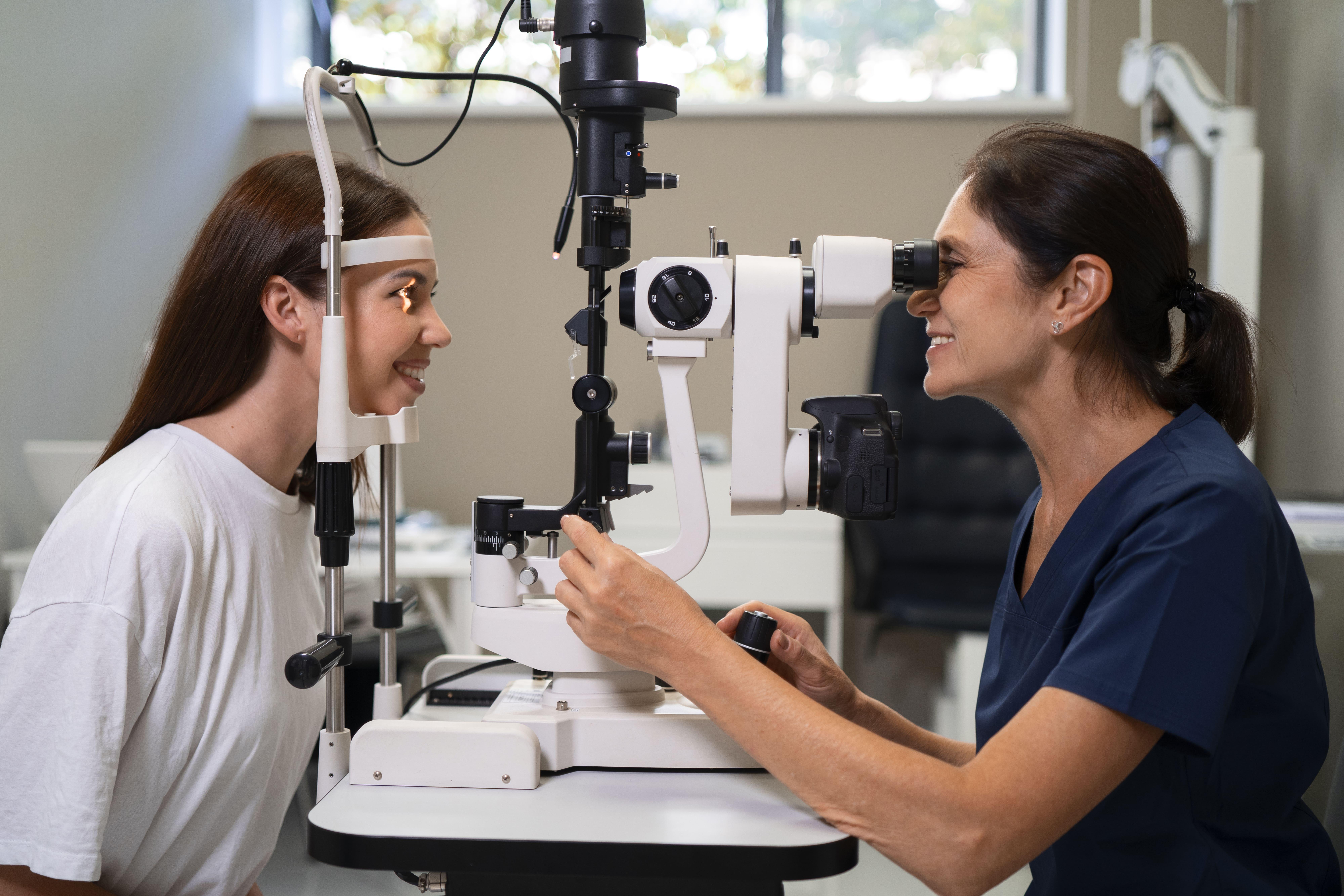
In ophthalmic practice, the fundus camera is used to capture detailed retinal images that help clinicians identify retinal degeneration, vascular abnormalities, optic nerve changes, retinal swelling, and subtle structural disorders during routine retinal health monitoring procedures.
Accurate retinal documentation supports earlier intervention and improves long-term patient management strategies.
Many retinal conditions develop gradually and may not immediately affect vision. High-resolution retinal imaging allows clinicians to identify early abnormalities before severe complications occur.
Early retinal findings may include:
Small retinal hemorrhages
Mild retinal swelling
Microvascular irregularities
Pigment abnormalities
Early optic nerve changes
Early identification of these abnormalities improves treatment opportunities and helps preserve long-term retinal health.
Many retinal diseases require continuous observation because disease progression may occur slowly over time. Retinal imaging allows clinicians to compare images from different visits and evaluate subtle retinal changes more consistently.
Long-term monitoring supports:
Assessment of retinal stability
Detection of disease progression
Evaluation of treatment effectiveness
Improved clinical management strategies
Maintaining detailed retinal image records strengthens continuity of care and improves overall ophthalmic evaluation.
Improving Diagnostic Accuracy
High-quality retinal imaging improves diagnostic precision by allowing clinicians to evaluate retinal structures in exceptional detail. Enhanced visualization supports more reliable identification of abnormalities and improves differentiation between retinal disorders.
Improved diagnostic accuracy supports:
Better treatment planning
Reliable disease classification
Improved patient communication
Consistent retinal documentation
This level of precision contributes significantly to modern preventive ophthalmic care.
The quality of retinal monitoring depends greatly on the imaging systems used within ophthalmic clinics and diagnostic centers. High-performance retinal imaging equipment provides clearer visualization, enhanced image contrast, and more reliable diagnostic performance during retinal examination procedures.
Matronix Optotechnik offers advanced ophthalmic imaging solutions designed to support detailed retinal evaluation and efficient clinical workflows. Their systems are developed with modern optical technology that enables clinicians to achieve accurate retinal visualization, reliable image documentation, and improved diagnostic confidence during routine retinal assessment procedures.
Modern retinal imaging systems also improve workflow efficiency within ophthalmic practices. High-resolution retinal images can be captured quickly while maintaining excellent image quality and diagnostic detail.
Efficient imaging systems support:
Faster patient assessment
Better clinical documentation
Improved communication between specialists
Streamlined retinal screening procedures
These workflow improvements help clinics provide efficient and reliable ophthalmic care services for patients requiring long-term retinal monitoring.
Technological advancements continue improving retinal imaging capabilities and ophthalmic diagnostics. Artificial intelligence, automated image analysis, and enhanced digital imaging systems are helping clinicians identify retinal abnormalities with greater speed and precision.
Future developments are expected to improve:
Early disease detection
Diagnostic accuracy
Long-term retinal monitoring
Preventive ophthalmic care strategies
These innovations will continue strengthening retinal health monitoring and improving visual care outcomes worldwide.
Routine retinal health monitoring is essential for preserving visual function and identifying retinal abnormalities before severe complications occur. Advanced retinal imaging technologies have transformed ophthalmic diagnostics by enabling detailed retinal visualization, accurate image documentation, and improved long-term disease monitoring.
With reliable imaging systems and continued technological advancement, clinicians can improve diagnostic precision, strengthen preventive ophthalmic care strategies, and provide better visual care outcomes for patients undergoing routine retinal evaluation.